


I. INTRODUCTION
To give spatial context, specifically for entire cells, Biological imaging methods navigated to 3D in place of 2D via tomography or imaging. In biological analysis of volume, voxels are used to denote the data as a 3D grid and every voxel denoted as a consumption of few probe i.e. electrons or X-rays at that point. In the case of cryo-immobilized samples and highly difficult datasets proceeding these volumes were hard due to their low contrast and low signal-to-noise ratio [1]. Gradient boosting is a highly matured algorithm to update the boosting classifiers online and frequently enhances the exactness of the segmentation [5]. In order to manage the labeling uncertainty for semi-supervised learning and it explains the online gradient boosting technique which depends on the various instance learning. Weak classifiers in boosting were usually easy linear functions but decision trees were at time utilized for good generalization [6]; the output of the classifier is termed as gradient boosting decision tree.
Manuscript published on 30 August 2019.
*Correspondence Author(s)
Dr. Bhawna Nigam, Assistant Professor, Department of Information
Technology, Institute of Engineering & Technology, Devi Ahilya
University, Indore, India ,
Ramakrishnan M Ramanathaiah, Director-SAP Practice-Analytics
Big-Data-Cloud, Miracle software systems, USA.
Dr. Basavaprasad B., Assistant Professor, Govt. First Grade College,
Raichur, India ,
© The Authors. Published by Blue Eyes Intelligence Engineering and
Sciences Publication (BEIESP). This is an open access article under the
CC-BY-NC-ND license http://creativecommons.org/licenses/by-nc-nd/4.0/
II. RELATED WORK
The segmentation is acquired by segregating the pixels into various pixel classes. These classes are denoted by multi-variant Gaussian distributions. So, the only axiom regarding the nature of the features is that some added ingredients for the Gaussian noise model which is appropriate to explain the feature distribution which is own by the provided class. Here, it utilizes the perceptually uniform color values as color features and a set of Gabor filters as texture features. Gaussian parameters may be estimated by utilizing training data set or from the input image. It gives a parameter estimation method by utilizing the Expectation Maximization (EM) algorithm. Experimental results are given to explain the execution of the method on both synthetic and natural color images. In [10] Holtzman et al (2003) utilizes the hierarchical method in order to drew-out more object from the image. In every stage it selects one sub region that adds more than one object, and it separates it into two sub regions. For the provided image, it enforces the segmentation algorithm. Finally, in this stage, it has an external layer which explains the edges of the segmented object. If in case further segmentation is required, then it manually select the area created from the earlier step and it enforce the segmentation algorithm again specifically to the selected area. The same way, it separates the appealing part of the image. In [11] Alihodzic et al (2013) proposed Bat Algorithm (BA) is a biological algorithm, advanced and BA is defined as highly effective. BA depends on echo locations nature for multilevel thresholds for choosing, which utilizes the maximum entropy criterion. The experimental results explain the BA algorithm which search for multiple thresholds and it is very close to the optimal ones that are defined by the exhaustive search method. When distinguished with the current algorithms, the BA algorithm’s Excellency is exceeded. This algorithm is to explain the possibility of BA method for multilevel threshold. Along with this, it gives a new option to the traditional methods because of its modesty and effectiveness.
III. PROPOSED METHODOLOGY
In the proposed methodology, improved bat algorithm using super-region volume segmentation for medical images is proposed. The general block diagram is explained in Fig 1.
Improved Bat Algorithm using Super-Region Volume Segmentation for Medical Images
A. Data Collection and Preprocessing
We used two samples neuronal-like mammalian cell line and Trypanosoma brucei procyclic cells for testing to segmentation with improved SuRVoS. The neuronal-like mammalian cells are implemented onto gold finder Transmission Electron Microscopy (TEM) grids. The Trypanosoma brucei procyclic cells were experienced and fixed priory in glutaraldehyde to include 200nm gold fiducials, pipetting onto copper TEM grids with lacey carbon support and cryo-immobilization using a Mark IV (FEI) plunge freezer. The data pre-processing step links de-noising and textural feature extraction methods to improve the attitude of the data and create future classification easier. De-noising adds Gaussian and Total Variation (TV) filters. While Gaussian filters generally creates over-smoothed results, TV methods maintain strong volume edges. It leads to split the volume in piece-wise smooth regions. Hence, a Gaussian-denoized volume is better to adapt for super-region extraction, while TV de-noising offers good execution to extract the features for classification.
B. SuRVoS framework
At present, the MRC file format (.mrc) and HDF5 file format (.h5, .hdf5 is favored by SuRVoS. After loading the data into the tool, the variations can be selected mechanically and adapted manually. User interface of the workbench adds three various regions, the Visualization Pane, the Plugins and the Tool Column. Utilizing the Plugins and evaluated in the Visualizations Pane can select and enforce the Parameters, while the Tool Column houses shortcuts were utilized in tools often. Multiple RoIs can be generated in design of the bounding cuboids. Remaining actions of tool can be restricted to the entitled RoI.
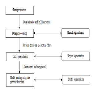
Fig. 1 : Block Diagram of the Proposed System
This permits the user to choose the RoI to validate the de-noising and textural features in a smaller area before extending then to the entire volume or preferably it permits the user to restrict the automatic segmentation to a particular Region of Interest (RoI).In the proposed work, new formulation is performed with the assistance of intelligent behavior. In this work it proceeds the procedure of Bat Algorithm based on MRF formulation. Nevertheless, the proposed algorithm resolves the energy function into an image-labeling issue to control the label consistency conditions.
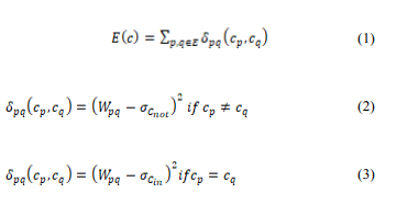
The energy function can be resolved by bat algorithm. This BA enhances the label MRFs by continuously extend the individual label by utilizing the graph cut. Graph cut can identify the optimal expansion if the expansion is sub modular.
IV. EXPERIMENTAL RESULT
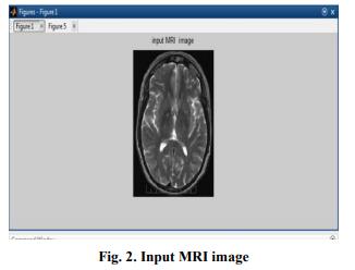
For the evaluation of the proposed tumor classification, the DICOM brain images are utilized. A total of 760 images are utilized of which 500 are for training and 260 are for testing. In the training images, 272 are tumor and 228 are non-tumor images while in testing images 150 are tumor and 110 are non-tumor. The size of the image is 256×256 pixels have a total of 65536 pixels size with resolution of 96ppi.The 3D data are exotic into SuRVoS, where the contrast is adapted and the volume is pruned to the appropriate area. In the pre-processing tab, the data is scaled and suitable supervoxels and megavoxels are detected. The experiments were proceeded through MATLAB. Matlab is a programming language which can be utilized for a broad array of numerical and computing applications. It measures the efficiency of the image compression performance. The computation of the proposed algorithm is distinguished with the current SuRVoS method. To the measure the implementation of the current and the proposed approaches, various parameters were utilized like MSE, PSNR, correlation, accuracy, precision and, recall.
V. CONCLUSION
The proposed method models are a combination of the initialization techniques to segment structures in volume data. The user interface is to mutually segment huge 3-D volumes. Super-Regions (megavoxels and supervoxels) minimize the manual segmentation and eliminate manual delineation of boundaries, conceivably reduce the subjectiveness. Furthermore, the proposed method gives a broad variety of de-noising and textural filters which is tuned to every sample, and utilized to guide the machine learning model to expand user’s manual segmentation to the volume of remaining set. Megavoxels and Supervoxels were utilized in combination with the model training to segment the cytoplasm, nucleus, and extracellular area. Furthermore, to the above advantages by utilizing the features which is brought-in in this proposed method, user’s segmentation time is minimized by five times when compared with the manual segmentation. Along with this, these features provide a way for good and very quantitative utilization of the 3-D volume datasets in science related to biology. The improved bat algorithm is utilized to give an enhanced model to produce the great fitness function values. The results confirm the proposed system gives excellent performance output in terms visual perception of image segmentation.
REFERENCES
- Lucic, V., Rigort, A., Baumeister, W., 2013. Cryo-electron
tomography: the challenge of doing structural biology in situ. J. Cell
Biol. 202 (3) - Zhang, Kaihua, et al. “Active contours with selective local or global
segmentation: a new formulation and level set method.” Image and
Vision computing 28.4 (2010): 668-676. - T. Chan, L. Vese, Active contours without edges, IEEE Transaction on
Image Processing 10 (2) (2001) 266–277. - A. Bosch, A. Zisserman, and X. Munoz. Image classification using
random forests and ferns. In Proc. ICCV, 2007. - Son, Jeany, et al. “Tracking-by-segmentation with online gradient
boosting decision tree.” Proceedings of the IEEE International
Conference on Computer Vision. 2015. - J. H. Friedman. Greedy function approximation: A gradient boosting
machine. Annals of Statistics, 29:1189–1232, 2000. 2. - Tzotsos, Angelos, and Demetre Argialas. “Support vector machine
classification for object-based image analysis.” Object-Based Image
Analysis. Springer Berlin
Heidelberg, 2008. 663-677 - Kato, Zoltan, and Ting-Chuen Pong. “A Markov random field image
segmentation model for color textured images.” Image and Vision
Computing24.10 (2006): 1103-1114. - Ding, Lei, and Alper Yilmaz. “Interactive image segmentation using
probabilistic hypergraphs.” Pattern Recognition 43.5 (2010):
1863-1873. - Holtzman-Gazit, Michal, Dorith Goldsher, and Ron Kimmel.
“Hierarchical segmentation of thin structures in volumetric medical
images.” International Conference on Medical Image Computing and
Computer-Assisted Intervention. Springer Berlin Heidelberg, 2003. - Alihodzic, Adis, and Milan Tuba. “Bat algorithm for image
thresholding”, RRT, Informatics, Electronics and Signal Processing
(2013): 17-19. - Ayan Nigam, Bhawna Nigam, Chayan Bhaisare, Neeraj Arya,
Classifying the bugs using multi-class semi supervised support vector
machine, International Conference on Pattern Recognition, Informatics
and Medical Engineering (PRIME-2012) - M Niranjanamurthy, B Nigam, Nithya, S Jagannatha- “Analysis of
Blockchain technology: pros, cons and SWOT” Cluster Computing
The Journal of Networks, Software Tools and Applications ISSN:
1386-7857 (Print) 1573-7543 (Online)
https://doi.org/10.1007/s10586-018-2387-5 2018 - M Niranjanamurthy, D Chahar – “The study of e-commerce security
issues and solutions” International Journal of Advanced Research in
Computer and Communication Engineering Vol. 2, Issue 7, ISSN
(Online) : 2278-1021 P:1-12 June 2013 - Niranjanamurthy M, Bhawna Nigam, Niveditha N.M , Naresh E –
“Efficient Implementation of Refund Process in Online
ShoppingIndustry Internal Tool-OMS” International Journal of Recent
Technology and Engineering (IJRTE) ISSN: 2277-3878,
Volume-7Issue-6, March 2019
AUTHORS PROFILE
Dr. Bhawna Nigam received Ph.D. in Computer Engineering from Devi Ahilya University, Indore, M.P. in 2017, M.E. in Software Engineering with distinction from Institute of Engineering & Technology (IET), Devi Ahilya University in 2008 and B.E in 2003 from Institute of Engineering & Technology (IET), Devi Ahilya University, Indore. She is currently with Institute of Engineering and Technology (IET), Devi Ahilya University, Indore, India as Assistant Professor in Information Technology department. She is with Devi Ahilya University since 2007. Her area of interest are Machine Learning, Deep Learning, Data Mining, Big Data.She has published 20+ papers on these topics.
Ramakrishnan M Ramanathaiah, SAP Practice-Analytics-Big Data-Cloud, Miracle software systems, USA. is a senior solutions architect and Director with 17 years of industry experience, Ram is extremely well-versed architect capable of handling highly complex projects in Analytics, AI, Machine learning, Big Data and cloud. Ram is working in US for more than a decade, prior to that he worked in Singapore, Thailand for few years.
Dr. Basavaprasad B. working as Assistant Professor, Govt. First Grade College, Raichur, India. He received Ph.D. in Computer Engineering from Bharathiar University, India in 2019, M.Sc. in Computer Science from Gulbarga University in 2004. His area of interest are Digital Image Processing & Pattern Recognition, Software Engineering. He has published 20+ papers on these topics.

